Key points of this research result
- Newly produced proteins are folded in the endoplasmic reticulum (*1) and transported to the Golgi apparatus (*3) by transport capsules called COPII vesicles (*2). However, during times of stress, abnormal proteins increase, and their transport becomes a problem. Therefore, cells have a mechanism to prevent abnormal proteins from being released, but the details have not been clarified.
- When investigated in Saccharomyces cerevisiae (*4), it was found that the membrane lipid phosphatidic acid (PA) (*6) (PA) increases during endoplasmic reticulum stress (*5). This change occurs through gene expression.YIP3Increases the protein encoded by genes (*7). As a result, it was revealed that the accumulation of Sec16 (*8), which forms the basis of transport capsules, on membranes was suppressed, reducing protein transport.
- This study demonstrated how cells use membrane changes to adjust their logistics during times of stress. Endoplasmic reticulum stress is an important phenomenon involved in the pathology of various diseases such as neurodegenerative diseases, diabetes, and cancer. Elucidation of the stress response mechanism is expected to lead to the development of new treatments.
overview
Kazuki Hanaoka (graduate student), Mitsuki Nakazato (graduate student), Philipp Schlarmann (special foreign researcher), Koichi Funato (professor) from the Graduate School of Integrated Life Sciences, Hiroshima University, Muneyoshi Kanai (senior researcher) and Haruyuki Iedo (former department head) from the National Alcoholic Beverage Research Institute, Howard Riezman (professor) from the University of Geneva, and Manuel from the University of Seville. An international collaborative research group led by Professor Muñiz has revealed a new regulatory mechanism for how proteins are transported within cells.
Inside the cell, newly created proteins are first reformed in the endoplasmic reticulum, then transported to the Golgi apparatus, where they are finally delivered to where they are needed. This transport from the endoplasmic reticulum to the Golgi apparatus is carried out by small capsule-like structures surrounded by a lipid membrane called COPII vesicles.
However, when cells are stressed, proteins cannot be formed properly, and incomplete proteins accumulate in the endoplasmic reticulum. Cells have a mechanism that prevents these abnormal proteins from being released until they are repaired, but the detailed mechanism has not been clarified.
Through research using yeast, this research group discovered that the lipids in cell membranes change during times of stress, and this change triggers the suppression of protein transport. especially"YIP3We discovered that the gene ``'' plays an important role.
YIP3Yip3, a protein produced by , suppresses the function of Sec16, an important protein that serves as a "scaffold" when constructing transport capsules. This is thought to prevent capsule formation and reduce the movement of proteins from the endoplasmic reticulum to the Golgi apparatus.
These results revealed that the endoplasmic reticulum has a mechanism that senses changes in the state of its membrane and adjusts the flow of proteins when it receives stress. This mechanism is thought to be related to neurological diseases, diabetes, cancer, etc., and this result is expected to lead to the understanding of diseases and the development of new treatments.
This research was published in the international scientific journal "Nature Communications" on July 3, 2020 (Japan time).
Paper information
Publication magazine name: Nature Communications
Paper title: ER sensing of lipid metabolism drives PRA family-dependent regulation of COPII vesicle transport
Author names: Kazuki Hanaoka†, Mitsuki Nakazato†, Philipp Schlarmann†, Hiroki Nakamura, Mei Kato, Ryoko Ikema, Mizuki Iguchi, Katsuki Eto, Takefumi Karashima, Atsuko Ikeda, Yukari Yabuki, Javier Manzano-Lopez, Auxiliadora Aguilera-Romero, Susana Sabido-Bozo, Ana Maria Perez-Linero, Muneyoshi Kanai, Haruyuki Iefuji, Isabelle Riezman, Howard Riezman, Manuel Muñiz, Kouichi Funato*
(†Co-lead author)(*Corresponding author)
DOI: https://doi.org/10.1038/s41467-026-75057-x
background
In the cells that make up our bodies, life activities are maintained by transporting proteins and lipids to the correct locations. Among these, the endoplasmic reticulum is an important organelle (*9) in which proteins and lipids are produced. Proteins and lipids produced in the endoplasmic reticulum are wrapped in transport vesicles called COPII vesicles and transported to the Golgi apparatus, where they are then delivered to their respective destinations. This transport from the endoplasmic reticulum to the Golgi apparatus is the starting point for intracellular logistics and is a basic mechanism widely conserved in eukaryotes from yeast to humans.
COPII vesicles are formed in a specific region called the ER exit site (ERES) (*10) on the endoplasmic reticulum membrane. In ERES, a group of proteins necessary for COPII vesicle formation assemble, and a protein called Sec16 acts as a scaffold to support the assembly, resulting in vesicle formation. In other words, ERES is the "shipping port" that initiates transport from the endoplasmic reticulum, and Sec16 plays an important role as the "foundation" for creating that shipping port.
On the other hand, the endoplasmic reticulum is not always maintained in a constant state. When cells are exposed to stress, the normal folding of proteins is disrupted, resulting in abnormal proteins that accumulate in the endoplasmic reticulum. This accumulation is thought to be due to a so-called quality control mechanism in the endoplasmic reticulum that prevents abnormal proteins from moving elsewhere until they are properly repaired. However, how endoplasmic reticulum stress is transmitted to inhibit COPII vesicle formation has not been fully elucidated.
Contents of research results
This research group used the model organism Saccharomyces cerevisiae to analyze how protein transport by COPII vesicles is regulated during endoplasmic reticulum stress.
First, we found that the amount of phosphatidic acid (PA), a type of phospholipid, increased in cells subjected to endoplasmic reticulum stress (Figure 1A). PA is known as a lipid that regulates the function of the transcription factors Ino2/Ino4 by controlling the localization of the transcription factor Opi1 on the endoplasmic reticulum membrane. Therefore, this research group searched for factors that suppress COPII vesicle transport downstream of the PA–Opi1–Ino2/Ino4 transcriptional control system (*11).
As a result of extensive genetic analysis, expression is regulated downstream of Ino2/Ino4 and has been identified as a new factor regulating COPII vesicle transport.YIP3We have identified the gene.YIP3The Yip3 protein encoded by was found to act as a factor that suppresses COPII vesicular transport. Indeed, we observed that the amount of Yip3 protein increased upon induction of endoplasmic reticulum stress (Figure 1B).
Next, we investigated how Yip3 suppresses COPII vesicle formation. COPII vesicles are formed with ERES on the endoplasmic reticulum membrane. In ERES, Sec16 acts as a "base" that gathers the proteins necessary for COPII vesicle formation. In this study, we investigated the extent to which Sec16 gathers on the endoplasmic reticulum membrane using fluorescence microscopy. As a result, we found that Sec16 accumulation in ERES decreased during endoplasmic reticulum stress (Figure 1C).
From the above results, during endoplasmic reticulum stress, the amount of PA in the endoplasmic reticulum membrane increases andYIP3It was revealed that gene expression was promoted and the increased Yip3 protein inhibited the formation of COPII vesicles by suppressing the accumulation of Sec16 in ERES (Figure 2).
This study demonstrated that the endoplasmic reticulum has a mechanism that uses the state of its own membrane lipids as cues to regulate the amount of intracellular shipment through transcription.
Future developments
This study revealed that phosphatidic acid (PA) levels in the ER membrane increase during ER stress, and this information leads to suppression of COPII vesicle formation through transcription.
On the other hand, in this study, the localization of Yip3 and Sec16 within the cell did not completely match, and no clear physical interaction between the two was confirmed. This suggests that Yip3 does not directly control Sec16, but may control the accumulation of Sec16 into ERES through another factor. In the future, it will be important to clarify which molecule Yip3 suppresses Sec16 accumulation through.
In addition, it was shown that downstream of the Opi1–Ino2/Ino4 transcriptional control system, there is a possibility that there is an unknown factor (XXX) other than Yip3 that controls the first step of COPII vesicle formation (Figure 2).Identification of these factors will greatly contribute to elucidating the entire transport control mechanism during endoplasmic reticulum stress.
Yip3 is a protein that belongs to the PRA family (*12), and this family is widely conserved from yeast to humans. Therefore, the mechanism clarified this time will provide important clues for understanding the protein transport control mechanism during endoplasmic reticulum stress in higher eukaryotes, including humans.
Endoplasmic reticulum stress is involved in the pathology of various diseases including neurodegenerative diseases, diabetes, and cancer. Based on these results, progress in elucidating the mechanism of COPII vesicle formation control and the endoplasmic reticulum stress response mechanism mediated by lipids and PRA family proteins is expected to greatly contribute to the understanding of the etiology and pathophysiology of various diseases based on secretion failure, breakdown of endoplasmic reticulum homeostasis, or abnormal lipid metabolism, and lead to the development of new therapeutic methods.
Reference materials
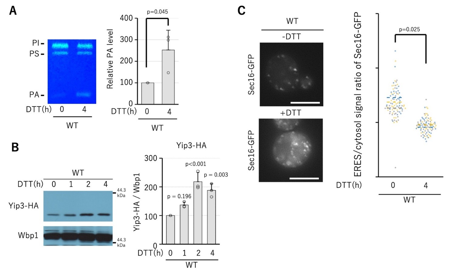
Figure 1. ER stress increases the amount of phosphatidic acid (PA) and Yip3 and reduces the accumulation of Sec16 in ERES.
A wild-type strain was treated with DTT (dithiothreitol), a reducing agent that causes endoplasmic reticulum stress, for 4 hours, and the amount of PA in the cells was analyzed by thin layer chromatography (TLC). As a result, it was confirmed that DTT treatment increased the amount of PA.
BYIP3Cells expressing Yip3-HA, an HA-tagged gene, were treated with DTT for 1, 2, and 4 hours, and the amount of Yip3-HA was analyzed by Western blotting. As a result, it was confirmed that the amount of Yip3-HA increased in a time-dependent manner due to endoplasmic reticulum stress.
C Cells expressing Sec16-GFP, which is a GFP-tagged Sec16, were treated with DTT for 4 hours, and the intracellular localization of Sec16-GFP was observed using a fluorescence microscope. In DTT-untreated cells, Sec16-GFP was observed in the form of dots (ER exit site) on the endoplasmic reticulum membrane, and the signal intensity ratio between the dots and the cytoplasm was high. On the other hand, in cells treated with DTT, the dot-like signal of Sec16-GFP decreased, a signal widely diffused in the cytoplasm was observed, and the signal intensity ratio between the dots and the cytoplasm decreased. This result indicates that ER stress makes it difficult for Sec16, which serves as the "foundation" for COPII vesicle formation, to gather at ERES.
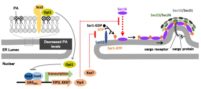
Figure 2. A model of the mechanism that senses phosphatidic acid (PA) levels in the endoplasmic reticulum membrane and inhibits COPII vesicle formation.
When PA levels in the endoplasmic reticulum membrane are low, Opi1 translocates into the nucleus and suppresses the transcriptional activity of Ino2/Ino4. the result,YIP3The expression of genes and unknown factors XXX decreases, and the suppression of COPII vesicle formation is released.
On the other hand, when PA levels in the ER membrane increase due to ER stress, Opi1 remains on the ER membrane and transcriptional activation by Ino2/Ino4 is maintained. the result,YIP3Gene expression increases, and Yip3 protein prevents the accumulation of the scaffolding protein Sec16 for COPII vesicle formation at the ERES. This prevents the "foundation" for COPII vesicles from being built and limits protein transport from the endoplasmic reticulum to the Golgi apparatus.
This study raised the possibility that, in addition to Yip3, there is an unknown factor XXX that controls the first step of COPII vesicle formation (binding of Sar1 to the endoplasmic reticulum membrane) downstream of Ino2/Ino4. These results suggest that the endoplasmic reticulum uses changes in PA levels to regulate COPII vesicle formation at multiple stages through transcription.
Explanation of terms
*1 Endoplasmic reticulum
One of the organelles that exists within eukaryotic cells. It is an important place where proteins and lipids are produced, and secretory proteins and membrane proteins in particular are transported to the Golgi apparatus after being folded normally in the endoplasmic reticulum.
*2 COPII vesicle
Transport vesicles that transport proteins and lipids from the endoplasmic reticulum to the Golgi apparatus. It is a small transport capsule wrapped in a lipid membrane that is formed when a part of the endoplasmic reticulum membrane is constricted, and functions as an intracellular transport vesicle that is widely conserved from yeast to humans.
*3 Golgi body
A cellular organelle that further modifies proteins and lipids transported from the endoplasmic reticulum and sorts them to appropriate locations inside and outside the cell. It plays a role like a collection and sorting center in intracellular logistics.
*4 Saccharomyces cerevisiae
A single-celled eukaryote that is also used to ferment bread and alcohol. They share many basic biological phenomena and mechanisms with humans, and are relatively easy to genetically manipulate and observe inside cells, so they are widely used as model organisms in life science research.
*5 Endoplasmic reticulum stress
Intracellular stress that occurs when proteins do not fold properly in the endoplasmic reticulum and abnormal proteins accumulate. Endoplasmic reticulum stress is known to be associated with various diseases such as neurodegenerative diseases, diabetes, and cancer.
*6 Phosphatidic acid (PA)
A type of phospholipid that makes up the membranes of cell membranes and organelles. It has a small hydrophilic head and a hydrophobic tail made of fatty acids, which have characteristics that affect the shape and bendability of the membrane. It not only serves as a material for membrane lipids, but also plays a role in intracellular information transmission and regulation of lipid metabolism. In this study, we showed that PA levels increase during endoplasmic reticulum stress and suppress COPII vesicle formation.
*7YIP3gene
A gene present in Saccharomyces cerevisiae that encodes the Yip3 protein, which belongs to the PRA family. Through this research,YIP3was identified as a new factor whose expression is regulated downstream of the Opi1–Ino2/Ino4 transcriptional control system and suppresses the formation of COPII vesicles by suppressing the accumulation of Sec16 in the ERES.
*8 Sec16
Scaffolding proteins required for COPII vesicle formation. It gathers at ERES on the endoplasmic reticulum membrane and plays a role like a “base” that gathers the proteins necessary to create COPII vesicles.
*9 Cell organelle
A structure that plays a specific function within a cell. Most exist as small compartments separated by lipid membranes, and include the endoplasmic reticulum, Golgi apparatus, mitochondria, vacuole, and nucleus. The vital activities of cells are maintained by each of them sharing their roles.
*10 ER exit site (ERES)
A specific region on the endoplasmic reticulum membrane where COPII vesicles are formed. At the "shipment port" where transport from the endoplasmic reticulum to the Golgi apparatus begins, proteins such as Sec16 gather to support the formation of COPII vesicles.
*11 Opi1–Ino2/Ino4 transcription control system
In yeast, a transcriptional control system that regulates gene expression according to the intracellular metabolic state. Ino2/Ino4 functions as a transcription factor that promotes gene expression, and Opi1 functions as a transcriptional regulator that suppresses this function. Opi1 changes its localization depending on the amount of PA on the endoplasmic reticulum membrane, so cells sense changes in the amount of PA on the endoplasmic reticulum membrane through this mechanism. In this study, we demonstrated that this transcriptional control systemYIPIt was shown to control the expression of 3 genes and lead to the regulation of COPII vesicle formation.
*12 PRA family
A family of membrane proteins that is widely conserved from yeast to humans. PRA is a name derived from Prenylated Rab Acceptor, and is known as a group of proteins involved in intracellular transport. Yip3 is a PRA family protein of Saccharomyces cerevisiae and was identified in this study as a factor that suppresses COPII vesicle formation.
- Press release materials (434.11 KB)
- Publication journal (Nature Communications)
- Hiroshima University Researcher Guidebook (Professor Koichi Funato)
(regarding research)
Food Life Science Program, Graduate School of Integrated Life Sciences, Hiroshima University
Professor Koichi Funato
Tel: 082-424-7923
E-mail: kfunato*hiroshima-u.ac.jp (Please change * to half-width @)
(Regarding reporting)
Hiroshima University Public Relations Office
E-mail: koho*office.hiroshima-u.ac.jp (Please change * to half-width @)