Key points of the presentation
- The model organism nematode (C. elegans) has been developed to perform infrared imaging while alive.
- Visualize the distribution of biomolecules such as proteins and lipids without labeling
- Visualizing differences in protein shapes (structures) in vivo by analyzing infrared spectra
- In the future, it is expected to be applied to research on diseases involving protein aggregation, such as Alzheimer's disease.
overview
名古屋工業大学工学専攻生命・物質化学プログラムの脇田 和佳氏(博士前期課程2A research group led by Assistant Professor Tatsuro Nishikino and Associate Professor Yuji Furuya of the Department of Life Science and Applied Chemistry, Professor Mitsumasa Koyanagi and Professor Akihisa Terakita of the Graduate School of Science, Osaka Public University, is working on C. elegans (2013), which is widely used as a model organism.C. elegans)を生きたまま赤外イメージングし、タンパク質の構造分布を可視化する技術を開発しました。
The research group used heavy water to suppress the influence of water that interferes with infrared observation, and also optimized the conditions for temporarily stopping nematode movement using sodium azide. As a result, we succeeded in obtaining high-quality infrared images from living nematodes. Furthermore, by second-order differential analysis of the obtained infrared absorption spectra, it was possible to identify typical structures that form proteins.αHelix andβWe visualized the differences in structures called sheets. As a result, in the head of the nematode, compared to other parts,βIt became clear that the proportion of sheet structures was low.
This research provides a new technological platform for visualizing protein structures in vivo without labeling with fluorescent dyes, and is expected to be applied to research on neurodegenerative diseases such as Alzheimer's disease in the future.
The results of this research are2026year8月26American Chemical Society open access journalACS OmegaPublished in
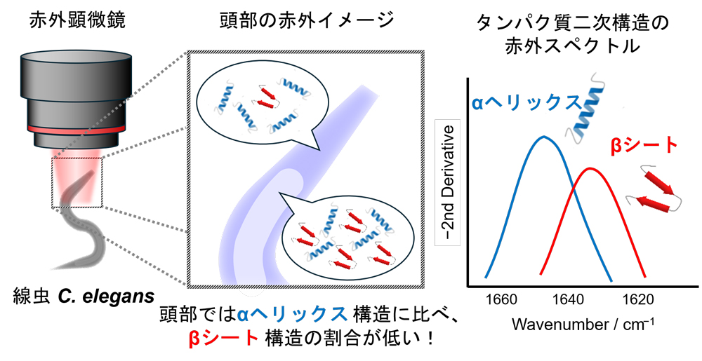 Research overview diagram
Magazine information
【発表雑誌】 ACS Omega
[Paper title] Minimally Invasive Infrared Imaging ofCaenorhabditis elegansEnabled by Reversible Chemical Immobilization and Second-Derivative Spectral Mapping
【著者】 Nodoka Wakita, Akiko Ito, Ryu Sato, Akihisa Terakita, Tatsuro Nishikino, Mitsumasa Koyanagi, and Yuji Furutani* *責任著者
[Publication URL]
https://pubs.acs.org/doi/10.1021/acsomega.6c03485
https://doi.org/10.1021/acsomega.6c03485
Osaka Public University Graduate School of Science
教授 小柳 光正(こやなぎ みつまさ)
TEL: 06-6605-2583
E-mail:koyanagi[at]omu.ac.jp
※[at]を@に変更してください。
大阪公立大学 広報課
TEL:06-6967-1834
E-mail:koho-list[at]ml.omu.ac.jp
※[at]を@に変更してください。
Source: https://www.omu.ac.jp/info/research_news/entry-25635.html