August Paper of the Month: An artificial intelligence optimized hepatic differentiation unveils NR5A2 and AP-1 transcriptional regulation in hepatic maturation
Liver cells, or hepatocytes, are responsible for performing crucial functions like filtering toxins, breaking down fats, and processing drugs. However, real human liver cells do not multiply well in a petri dish. To address this, scientists guide stem cells through key developmental stages to create mature hepatocyte-like cells (HLCs):
- Stem Cells (hPSCs)
- Definitive Endoderm (DECs)
- Hepatic Progenitor Cells (HPCs)
- Mature Hepatocyte-Like Cells (HLCs)
Historically, tracking this multistep process was a major bottleneck because it is quite difficult to visually assess whether intermediate cells are healthy and on track.
Teaching AI to spot healthy cells
To solve this problem, PhD candidate Mo-Fan (Elena) Huang, mentored by Dung-Fang Lee, PhD, along with her team, built a machine-learning AI tool that analyzes microscopic images of liver progenitor cells (HPCs, the “teenager”" stage) to assess differentiation success. This work is now published in the Journal of Biological Chemistry (JBC). Huang is a student in the Genetics and Epigenetics Program at The University of Texas MD Anderson Cancer Center UTHealth Houston Graduate School of Biomedical Sciences, where Lee is a faculty member as well as associate professor of integrative biology and pharmacology at McGovern Medical School at UTHealth Houston.
“Our main question was how to better define and characterize the intermediate stages of hepatic differentiation,” Huang said. “We wanted to identify reliable molecular markers and key transcriptional regulators that drive the transition from pluripotent stem cells to mature hepatocytes. Understanding these stage-specific regulatory mechanisms could help improve the efficiency and reproducibility of in vitro liver differentiation.”
To train the model, the team input hundreds of standard microscope photos into the AI and categorized them as successful, failed, or undetermined. The AI learned subtle visual patterns and achieved ~97.8% accuracy in predicting cell fate. This means they were able to reduce human error while saving time and resources.
Proving lab-grown liver function
By using this AI quality-check to ensure only high-quality cells progressed to the final stage, the resulting liver cells performed remarkably well. More than 95% of the final cells produced albumin (ALB), synthesized urea, and processed drugs similarly to primary human donor liver cells. Furthermore, when transplanted into preclinical models, the cells survived and matured into functional liver tissue.
Cracking the genetic code of liver maturation
To understand the underlying biology, the team mapped gene activity across all four stages. Stem cell genes gradually turned off while liver metabolism genes turned on as cells matured.
Most importantly, researchers discovered that two key protein families, NR5A2 and AP-1, drive this transformation: Single-cell analysis confirmed that NR5A2 ramps up early during the immature progenitor stage, while AP-1 components (like JUN, JUNB, FOSL2) peak right in the final phase to drive maturation.
Confirming safety and genetic stability
Ensuring safety is a vital step before these cells can be used in medicine. Using whole-genome sequencing, Huang and her team showed that the 21-day differentiation process did not cause dangerous DNA mutations. Additionally, cancer risk tests found that the cells were not growing uncontrollably in dishes and formed no tumors in preclinical models, proving they are genetically stable and safe.
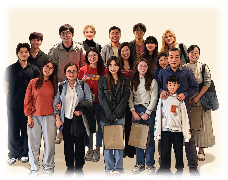
An illustration of Lee lab members.
Bringing the science full circle
“My favorite part was seeing the project come full circle,” Huang said. “We combined computational analyses to identify candidate regulators and then experimentally validated their functions, ultimately establishing an optimized differentiation protocol. It was especially rewarding to see our bioinformatics predictions confirmed through functional experiments.”
By combining AI visual tracking and mapping genetic changes, this team created a reliable roadmap for growing pure, safe liver cells. This breakthrough paves the way for superior drug testing, disease modeling, and future regenerative therapies for liver diseases.
“This work provides a framework for improving stem cell-derived hepatocyte differentiation by identifying key transcriptional regulators involved in hepatic maturation,” Huang concluded. “Our findings demonstrate how artificial intelligence-guided analyses, combined with experimental validation, can uncover regulatory networks that improve differentiation protocols. We hope this strategy will facilitate the generation of more mature and reproducible hepatocyte models for studying liver development, disease modeling, and regenerative medicine.”
UT MD Anderson Cancer Center UTHealth Houston Graduate School of Biomedical Science’s Paper of the Month is a collaborative effort led by PhD candidate Mirrah Bashir (author of August’s summary); Shelli Manning, communications manager; and Lauren Nguyen, communications assistant. Oversight is provided by Francesca Cole, PhD, associate dean for Academic Affairs at UT MD Anderson UTHealth Houston Graduate School. Students are trained to summarize fellow student-authored scientific articles about their biomedical science research and the innovative methods and discoveries they are uncovering. The Paper of the Month editorial team includes students Amanda Warner, Chae Yun Cho, Sheighlah McManus, Sarah Schneider, Trisha Wathan, Anna Debruine, and Trithi Sunder.