Researcher information
Researcher name
Kyoji Sumigama
overview
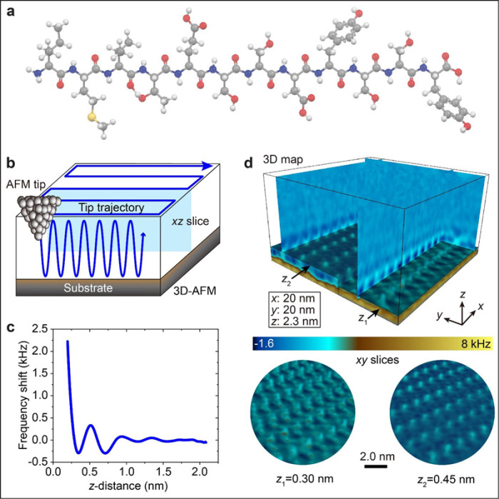
Professor Tsuyoshi Fukuma of Kanazawa University Nano Life Science Institute (WPI-NanoLSI), Specially Appointed Assistant Professor Ayhan Yurtsebel, Special Lecturer Kyoji Sumigata of Kyoto University Graduate School of Life Sciences/Kanazawa University Nano Life Science Institute, Mehmet of the University of Washington, USA An international collaborative research group led by Professor Sarikaya and colleagues has succeeded in directly visualizing for the first time in the world that water molecules surrounding protein surfaces form highly ordered three-dimensional structures according to amino acid sequences.
In recent years, advances in protein structure prediction technology using artificial intelligence (AI) have made it possible to predict the three-dimensional structure of proteins with high accuracy. However, even if the amino acid sequence and three-dimensional structure are known, it is still difficult to accurately predict its function, intermolecular interactions, and behavior in vivo. In this study, we used state-of-the-art three-dimensional atomic force microscopy (3D-AFM) to observe the hydration structure around peptide aggregates with subnanometer resolution. The results revealed that water is not just a solvent, but forms a multilayered and regular structure depending on the properties of hydrophobicity, polarity, aromaticity, and charged amino acid residues.
Furthermore, this research group considered the protein body and the unique hydration structure formed around it as an inseparable, integrated structure, and proposed a new concept called ``Protein Superstructure.''
The results of this research will greatly advance the understanding of protein functions and are expected to have applications in a wide range of fields such as drug discovery, protein engineering, and biomaterial development.
The results of this research were published in the online version (open access) of the international academic journal "Nature Communications" on August 26, 2026.
Source: https://www.kyoto-u.ac.jp/ja/research-news/2026-08-27-2