Toshifumi Shibata, Lecturer in the Department of Bacteriology, Tottori University School of Medicine (Global Health Research Center for Avian Infectious Diseases, Pathology Research Division), a joint research group from Okinawa Institute of Science and Technology Graduate University (OIST), Hiroshima University, and Nagasaki University, have announced thatBacteria that cause periodontal diseasePorphyromonas gingivalisFor the first time in the world, we have revealed the three-dimensional structure of the attachment organ "Mfa fimbriae" that plays an important role in the formation of dental plaque when Porphyromonas gingivalis (Porphyromonas gingivalis) infects humans.So I would like to inform you.
overview
Gingivalis bacteria has been linked not only to periodontal disease but also to a variety of other diseases, including aspiration pneumonia and Alzheimer's disease. However, the details of the infection mechanism are largely unknown, and there is no way to completely eliminate the bacteria.
This time, the research group has revealed for the first time in the world the actual structure of the ``Mfa pili'' that B. gingivalis uses to attach to human tissues and other bacteria. In addition to understanding how Mfa fimbriae are formed, we also found out at the molecular level that they have a special structure that allows them to escape from human immunity, and how they bind to streptococci in the oral cavity.
This research will enable us to understand at the molecular level how B. gingivalis uses Mfa fimbriae to attach to and infect human tissues. In the future, it is expected that this will lead to the development of new periodontal disease prevention and treatment methods that target Mfa fimbriae.
The results of this research were published online in the international academic journal "Communications Biology" on June 24, 2026.
Key points of this research result
- Cryo-electron microscopy revealed the three-dimensional structure of Mfa fimbriae, an attachment organ that functions as the "arms" used by Bacillus gingivalis, the bacteria that causes periodontal disease, for infection.*1revealed it at the atomic level for the first time in the world.
- Mfa fimbriae are proteases produced by bacteria.*2We revealed that the strands are assembled by a ``protease-dependent strand exchange mechanism'' that starts with cleavage by .
- We discovered that Mfa fimbriae have acquired a characteristic structure that utilizes calcium ions to escape from human immunity, and revealed a new immune evasion mechanism in P. gingivalis.
- By reproducing and visualizing the binding mode of Mfa fimbriae and oral pathogenic streptococci through computer simulation, we investigated the effects of bacterial aggregation and biofilms.*3We have clarified part of the (dental plaque) formation mechanism.
- The three-dimensional structure of Mfa fimbriae can be used as a target structure (template for drug discovery) for the development of drugs that inhibit the attachment and infection of periodontal disease bacteria, and is expected to lead to the development of new methods for preventing and treating periodontal disease.
background
Periodontal disease (periodontal pyorrhea) was recognized in the Guinness Book of Records in 2001 as the most prevalent disease in the world, and in Japan, approximately 80% of adults over the age of 30 are either affected or at risk of developing it.
Periodontal disease is an infectious disease in which Gingivalis bacteria that has invaded periodontal pockets adheres to tissues and forms a biofilm (dental plaque) in which multiple bacteria gather, causing chronic inflammation of the gums. As it progresses, the bone that supports the teeth is destroyed and the teeth fall out. Furthermore, C. gingivalis can invade the lungs from the oral cavity and cause aspiration pneumonia, or enter the bloodstream and spread throughout the body, causing or worsening various diseases such as diabetes, Alzheimer's disease, rheumatism, cerebral infarction, heart disease, and premature birth. On the other hand, there is still no method or drug to completely eliminate Gingivalis bacteria.
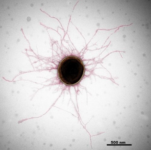
Figure 1: Electron microscopy image of B. gingivalis
Gingivalis is covered with thin filamentous structures called fimbriae (colored red by image processing). It infects humans using Mfa and Fim pili.
In order for pathogenic bacteria to infect, they must attach themselves to a host, such as a human or animal. Pili are filamentous attachment devices that act as "arms" for bacteria to bind to host tissues and other bacteria. Among them, the pili of B. gingivalis and its related species are classified as type 5 (type V) pili because they are constructed using a different formation mechanism from other bacterial pili (Reference 1). Gingivalis has two types of V-shaped fimbriae called "Fim fimbriae" and "Mfa fimbriae" (Figure 1), which each bind to specific oral proteins, immune-related molecules, and oral bacteria. In other words, it uses two types of fimbriae to infect and parasitize humans, causing disease.
In a previous study, the research group clarified the structure and formation mechanism of Fim pili (first arm) (Reference 2). By elucidating the properties of the Mfa fimbriae (the other arm) in this study, we have clarified the "full picture of both arms," which are responsible for the attachment and infection of M. gingivalis bacteria.
Contents of research results
The research group investigated Mfa1 pilin, a protein that composes Mfa fimbriae.*4polymerize in vitro*5We then performed structural analysis using cryo-electron microscopy. As a result, 3.0Å resolution*6We succeeded in determining the high-resolution three-dimensional structure of Mfa pili in its native state (Figure 2 ①). Based on this structural information and biochemical analysis results, we clarified the properties of Mfa fimbriae.
Cilia formation mechanism by “strand exchange”(Figure 2②)
When pilin Mfa1, a component of pili, is transported to the surface of B. gingivalis, the N-terminal region*7is cleaved by protease. Then, the internally folded β-strand in the C-terminal region undergoes a major structural change and is inverted and released. As a result, a hydrophobic groove is exposed inside the pilin. When the cleaved pilins approach each other, the β-strands of the adjacent pilins*8"Strand exchange" occurs where the pilins connect to the grooves, and the pilins connect with each other. It became clear that this reaction occurred continuously, resulting in the formation of Mfa fimbriae (Figure 2 ②A-F). This formation mechanism is also observed in Fim fimbriae, confirming that it is a universal formation principle common to V-type fimbriae.
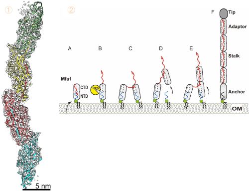
Figure 2: Mfa pili structure and assembly mechanism model by strand exchange
① Three-dimensional structure of Mfa fimbriae revealed by cryo-electron microscopy analysis. The Mfa1 pilin molecule binds to its neighboring pilin like an extended arm. ②Mfa pili construction model. After Mfa1 is transported to the surface of the bacterial cell as a membrane protein, a portion of the NTD is cleaved by the protease RgpB. This causes the C-end strand (red arrow) to flip (A-B). When the cleaved pilins approach each other, they join together at the C-terminal strand (C-D). This reaction is repeated and the pili elongates at the root and is constructed (E). Mfa1 constitutes Stalk, and other fimbriae include Tip, Adaptor, and Anchor (F).
Immune evasion mechanism of Mfa fimbriae by calcium binding
Structural analysis revealed that metal ions exist inside the Mfa1 molecule (Figure 3 ①). Further elemental analysis using ICP-AES (Inductively Coupled Plasma Emission Spectroscopy) determined that this metal ion was calcium. Even when the four amino acids that retain calcium were mutated to prevent calcium ion binding, pili formed normally (Figure 3 ②), indicating that calcium ion binding was not involved in pili construction. On the other hand, when this mutant fimbriae was applied to cultured human cells, it stimulated the production of IL-6, an inflammatory cytokine involved in the immune response.*9(Figure 3 ③). This indicates that the mutant Mfa fimbriae, in which the structure of the calcium ion binding region has been changed, are more easily recognized by the human immune system. From this, it can be inferred that M. gingivalis has incorporated calcium ions into its Mfa fimbriae during the evolutionary process in order to evade host immunity.
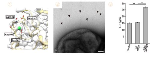
Figure 3: Calcium ion binding involved in immune evasion of Mfa fimbriae
① Calcium ions (green spheres) are bound to the loop structure containing aspartic acid (Asp), asparagine (Asn), and glutamic acid (Glu) in Mfa1. ② Mutant Mfa1 that does not bind calcium also polymerizes normally, forming fimbriae on the bacterial cells (arrow). ③ Calcium-binding mutant Mfa fimbriae (rMfa1DDNE/A) had higher immune induction to human cultured cells and increased IL-6 production compared to the untreated group (Control) and calcium ion-binding wild-type Mfa fimbriae (WTrMfa1). Wild-type fimbriae evaded immunity and produced the same amount of IL-6 as the untreated group.
Binding mode with oral streptococci
Mfa fimbriae are streptococci in the oral cavityStreptococcus gordoniiIt binds to the SspB protein on the surface of bacterial cells and causes co-aggregation between bacteria. This research has revealed the three-dimensional structure of the SspB binding region on the surface of Mfa pili, making it possible to analyze intermolecular interactions with high precision using computer simulations. This allowed us to reproduce and visualize the binding between Mfa pili and SspB.
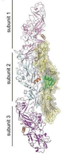
Figure 4: Simulation of binding of Mfa fimbriae to SspB of oral streptococci.
The binding between the determined Mfa fimbriae structure (three-molecule bond) and the previously elucidated SspB structure (yellow) was reproduced by computer simulation. The ⍺ helix on SspB, which is important for Mfa fimbriae binding, is highlighted in green.
Future developments and significance
These results will enable us to understand at the molecular level how B. gingivalis attaches to and infects humans via fimbriae, and how bacteria bind together to form biofilms. In addition, in recent drug discovery research, ``in silico drug discovery,'' which uses computers to virtually design, search, and narrow down compounds that bind to target proteins that cause diseases, has played an important role. To this end, high-resolution 3D structural information of the target molecule in its native state is essential. The detailed three-dimensional structure of Mfa fimbriae obtained in this study can be used as a drug discovery target structure (template for drug design) for designing molecules that inhibit the attachment and infection of M. gingivalis, and is expected to serve as the basis for the development of new periodontal disease prevention and treatment drugs. These efforts are expected to greatly contribute to maintaining human health by overcoming periodontal disease.
References
1. Xu, Shoji, Shibataet al. A Distinct Type of Pilus from the Human Microbiome.Cell 165, 690–703 (2016).
2. Shibataet alStructure of polymerized type V pilin reveals assembly mechanism involving protease-mediated strand exchange.Nat.Microbiol.5, 830–837 (2020)
Glossary
*1 Cryo-electron microscope (structural analysis):An electron microscope that can rapidly freeze biomolecules made of proteins and observe them under the low temperature of liquid nitrogen. The structure of molecules in their natural state can be visualized with high resolution. By processing the obtained image data, it is possible to construct and analyze the three-dimensional structure of molecules.
*2 Protease:An enzyme that cuts proteins. Pili polymerization involves a protease called gingipain RgpB secreted by B. gingivalis. Gingipain also plays a role as a pathogenic factor that degrades periodontal tissues and blood.
*3 Biofilm:Aggregates formed by bacteria adhering to surfaces. Bacteria create a sticky substance to cover themselves, which makes them resistant to drugs and immune systems, making them difficult to remove. Dental plaque is a typical example of oral biofilm.
*4 Pirin:A general term for proteins that are components of fimbriae.
*5 Polymerization:When many proteins combine to form a large structure. Mfa fimbriae are linked and elongated by the Mfa1 pilin protein.
*6 Resolution (Å Angstrom):An index that shows how detailed the structure can be seen through structural analysis. 1 Å is 1/100 millionth of a centimeter. The smaller the number, the more detailed the structure. With a resolution of 3 Å, the amino acids that make up proteins can be visualized, making it possible to understand the presence of metal ions and the bonding patterns between molecules with near-atomic-level precision.
*7 N end, C end (area):Proteins are made up of amino acids linked together, and the starting part is called the N-terminus and the ending part is called the C-terminus.
*8 β strand:One of the backbone structures (secondary structures) of proteins. A string-like structure consisting of 3 to 10 amino acids.
*9 Inflammatory cytokine IL-6:A type of information transmission protein that is secreted by immune cells and causes or enhances inflammatory responses. It is produced when bacterial or viral infections, tissue damage, etc. occur, and has the function of activating immune cells and protecting the body.
Paper information
●Title: Cryo-EM structure of the native assembled Mfa type V pilus from the periodontal pathogen Porphyromonas gingivalis
●Author: Satoshi Shibata*, Hideyuki Matsunami, Kazuhisa Ouhara, Yuri Taniguchi, Makoto Tokoro Schreiber, Alejandro Villar-Brillones, Koji Nakayama, Mikio Shoji and Matthias Wolf* (*Corresponding author)
●Publication journal: Communications Biology
●DOI: 10.1038/s42003-026-10515-2
Research support
This research was supported by Japan Society for the Promotion of Science (JSPS) Scientific Research Grants JP19K10083, JP23K06530, JP17K07318, JP20K06581 and Japan Agency for Medical Research and Development (AMED) JP25ama121037.
- Press release materials (860.32 KB)
- Publication journal (Communications Biology)
- Hiroshima University Researcher Guidebook (Lecturer Kazuhisa Ohara)
Department of Bacteriology, Department of Infection Control, Faculty of Medicine, Tottori University
Lecturer: Toshifumi Shibata
TEL: 0859-38-7580
E-mail: sshibata*tottori-u.ac.jp
Department of Emerging Infectious Diseases, Graduate School of Biomedical Sciences, Nagasaki University
Professor Mariko Naito
TEL: 095-819-7648
E-mail: mnaito*nagasaki-u.ac.jp
Hiroshima University Graduate School of Medical Sciences Laboratory of Periodontal Pathology
Lecturer: Kazuhisa Ohara
TEL: 082-257-5663
E-mail: kouhara*hiroshima-u.ac.jp
Tottori University Yonago Area Administration Department, General Affairs Division, Public Relations Section
TEL: 0859-38-7037
FAX: 0859-38-7029
E-mail: me-kouhou*ml.adm.tottori-u.ac.jp
Nagasaki University Public Relations Strategy Headquarters
TEL: 095-819-2007
E-mail: kouhou*ml.nagasaki-u.ac.jp
Public Relations Group, General Affairs and Public Relations Department, Hiroshima University
TEL: 082-424-3701
E-mail: koho*office.hiroshima-u.ac.jp
(Please replace * with half-width @)