Atsushi Otsuka, Chief Professor, Department of Dermatology, Kindai University School of Medicine (Sakai City, Osaka Prefecture), Doctor of Dermatology, Kindai University Hospital (Sakai City, Osaka Prefecture), Doctoral Student, Department of Medicine, Kindai University Graduate School of Medicine A research group led by Norimi Iinuma, in collaboration with Kyoto University (Kyoto City, Kyoto Prefecture), Sapporo Medical University (Sapporo City, Hokkaido), and Hiroshima University (Higashihiroshima City, Hiroshima Prefecture), has developed artificial intelligence (AI) that can automatically distinguish eight types of pigmented skin lesions (spots, moles, skin cancer, etc.) from ordinary skin images taken with a regular camera.
Until now, image diagnosis using AI has been highly accurate, but the reason for its judgment has been a black box, which has been a barrier to its use in medical settings. The newly developed AI uses a heat map that uses color shading to show where in the skin photo the AI was focused on when making a diagnosis.*2It is possible for people to visually confirm the ``reasons'' that have been identified. This research is significant in that it not only achieved high diagnostic accuracy but also was able to "explain" the basis for the decision.
A paper on this matter was published on July 23, 2026 (Thursday) in JAAD International, an international academic journal of dermatology published by Elsevier in conjunction with the American Academy of Dermatology (AAD).
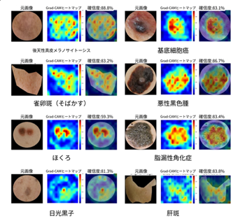
Heat map showing where AI looked and diagnosed
Key points of this case
- Developed AI to distinguish between 8 types of pigmented skin lesions and confirmed 95.9% diagnostic accuracy
- Visualize the basis of diagnosis by drawing a heat map of where the AI looked and diagnosed pigmented lesions
- The developed AI allows doctors to confirm the validity of the AI's judgments, so it is expected to be introduced in medical settings as a diagnostic support tool for pigmented skin lesions.
Background of the case
Pigmented skin lesions such as age spots, moles, and seborrheic keratosis are very common cases seen in dermatology practice. However, these pigmented lesions look similar to skin cancers such as malignant melanoma and basal cell carcinoma, and it is difficult for even specialists to diagnose them based on images alone. In order to reliably identify the*3Although this test is recommended, it is not always available in clinical settings other than dermatology, and it is not always possible to use it.
In recent years, research on skin image diagnosis using AI has progressed, and it has been reported that the accuracy is comparable to that of specialists. However, the more accurate AI is, the more complex its internal judgments become, and the ``black box problem'' in which it is difficult for humans to understand ``why a certain diagnosis was reached'' has become a barrier to its introduction into medical practice. Therefore, the research group developed an ``explainable AI'' that corresponds to pigmented lesions in Japanese people and can visually confirm the basis of diagnosis.*4We worked to realize this goal.
Contents of this case
The research group developed an AI that diagnoses eight types of pigmented skin lesions (acquired dermal melanocytosis, basal cell carcinoma, freckle, malignant melanoma, melasma, mole, seborrheic keratosis, senile pigment spot solar lentigo, and melasma) using 979 clinical photographs collected from multiple medical institutions in Japan. Ensemble learning that combines multiple deep learning models*5, and achieved a diagnostic accuracy of 95.9% on 196 test images that were not used for learning. Additionally, the system was able to correctly identify all 20 cases of malignant melanoma, which is a highly malignant cancer that is difficult to distinguish from a mole, in the test images.
Furthermore, in this study, Grad-CAM*6was used to visualize the areas on which AI made the diagnosis as a heat map. The results confirmed that AI diagnoses by focusing on the lesion itself, rather than the background, and showed that there are characteristics in the areas it focuses on depending on the type of lesion. This demonstrated not only high diagnostic accuracy but also the usefulness of ``explainable AI,'' which allows doctors to visually confirm the basis for a diagnosis.
This research is an internal validation of clinical photographs of Japanese people, and by proceeding with external validation, it is expected that the system will be used as a diagnostic support tool in areas where there are few dermatologists and in environments where dermoscopy cannot be used.
Paper published
Magazine: JAAD International (Impact factor: 6.5@2025)
Paper title: An Explainable Deep Learning Model Classifies Eight Categories of Pigmented Skin Lesions on Clinical Photographs: A Multicenter Retrospective Internal Validation Study in Japan
(Classification of pigmented skin lesions into 8 categories using clinical photographs using an explainable deep learning model: A multicenter retrospective internal validation study in Japan)
Author: Norimi Iinuma1, Kazuyasu Fujii2*, Chisa Nakajima2, Kenichiro Kasai3, Hiroyuki Irie4, Hitoshige Kintomo5, Shigeto Yanagihara5, Sayuri Sato6, Hisashi Uhara6, Fumiaki Takeda7, Yuichi Kimura8, Takashi Nagaoka9, Atsushi Otsuka2*Corresponding author
Affiliation: 1. Department of Dermatology, Kinki University Hospital, 2. Department of Dermatology, Kindai University School of Medicine, 3. Kasai Plastic Surgery Department, 4. Department of Dermatology, Kyoto University Graduate School of Medicine, 5. Kanetomo Dermatology Clinic, 6. Department of Dermatology, Sapporo Medical University, 7. Digital Manufacturing Education and Research Center, Hiroshima University. 8. Department of Informatics, Faculty of Informatics, Kindai University. 9. Department of Bioinformatics, Faculty of Physics and Engineering, Kinki University.
URL: https://doi.org/10.1016/j.jdin.2026.07.002
DOI: 10.1016/j.jdin.2026.07.002
Researcher's comments
Atsushi Otsuka (Atsushi Otsuka)
Affiliation: Kinki University School of Medicine, Department of Dermatology
Position: Chief Professor
Degree: Doctor (Medicine)
Comment: There are two types of pigmented lesions: benign and malignant, and distinguishing between them is very important clinically. This time, we have developed an AI that can highly accurately discriminate images taken with a common camera without using a special medical device called a dermatoscope. It is expected that its widespread use in medical settings will contribute to improving diagnostic accuracy in areas where there are no dermatologists.
Norimi Iinuma (Iinuma Kimi)
Affiliation: Kinki University Hospital Department of Dermatology
Position: Specialist doctor
Degree: Bachelor's (Medicine)
Comment: What we focused on most in this research was to show ``where the AI was looking and making that decision'' rather than the accuracy itself. The heat map showed us that the AI's line of sight was indeed capturing and capturing lesions, and that the points of view were different for each disease. We believe that AI that doctors can confirm the basis for can be safely delivered to clinical sites.
Glossary
*1 Pigmented skin lesions: A general term for lesions that appear as changes in color (pigment) on the skin, such as spots, moles, and skin cancer.
*2 Heat map: A typical visualization technology that uses color shading to indicate which areas are prioritized when AI classifies images.
*3 Dermoscopy: An examination method that uses a special magnifying glass to enlarge and observe the skin. Although it is useful for distinguishing pigmented lesions, it is often not usable outside of dermatology.
*4 Explainable AI: AI that can show in a form that humans can confirm the basis on which it makes decisions. In this study, we used a heat map that shows the places that the AI focused on in color.
*5 Ensemble learning: A machine learning method that increases diagnostic accuracy and stability by combining the prediction results of multiple AI models.
*6 Grad-CAM: Abbreviation for Gradient-weighted Class Activation Mapping. A technology in which image recognition AI uses a heat map to show "which part of the image it looked at to make a decision."
Where to distribute this document
Osaka Science and University Press Club, Education, Culture, Sports, Science and Technology Press Association, Science Press Association, Health, Labor and Welfare Press Association, Welfare Hibiya Club, Kyoto University Press Club, Hokkaido Education Press Club, Sakai City Government Press Club, Higashiosaka City Government Press Club
[Contact information regarding this matter]
Kinki University School of Medicine, Faculty of Nursing, Hospital Management Headquarters General Affairs and Public Relations Division Persons in charge: Morii, Hashimoto
TEL: 072-288-7222 FAX: 072-288-6303
E-mail: med.koho*itp.kindai.ac.jp
National University Corporation Kyoto University Public Relations Office International Public Relations Section
TEL: 075-753-5727 FAX: 075-753-2094
E-mail: comms*mail2.adm.kyoto-u.ac.jp
Hokkaido Public University Corporation Sapporo Medical University Corporate Planning Division Planning and Public Relations Section
TEL: 011-611-2111 (extension 21650)
E-mail: kouhou*sapmed.ac.jp
National University Corporation Hiroshima University Public Relations Group
TEL: 082-424-4383
E-mail: koho*office.hiroshima-u.ac.jp
(Please replace * with half-width @)