Key points of this research result
- We discovered that in human skin, before the amount of collagen (*1) and the shape of its fibers change, very small structural changes (damage) occur that are invisible to the naked eye.
- We have developed a new analysis method that combines multiple analysis techniques to examine not only the amount of collagen and the state of its fibers, but also the minute internal structure in the same sample.
- We demonstrated a new evaluation method that allows changes in collagen to be understood not only as a decrease in the amount of fibers but also as "changes in the internal structure."
- This new method is expected to be applied to the development of pharmaceuticals and cosmetics as a technology for investigating changes such as skin lesions and aging at an earlier stage.
overview
A research group led by Professor Katsuya Inoue of WPI-SKCM² (*2), Graduate School of Advanced Science and Engineering, Hiroshima University, has investigated the collagen contained in human skin in detail using a variety of analytical methods and discovered that even at a stage where there is almost no change in appearance, tiny structural changes have begun within the collagen.
In this study, we combined multiple analytical techniques to examine in detail the amount of collagen contained in human skin, the arrangement of fibers, and its internal structure using the same sample. The results revealed that even before visible changes such as a decrease in the amount of collagen or breakage of fibers appear, very small structural changes begin within the collagen.
This result demonstrated a new analysis method that allows for detailed evaluation of skin conditions, including changes in internal structure, which had previously been evaluated mainly based on changes in the amount of collagen and fiber shape. In the future, in addition to elucidating the mechanism of skin aging and disease, it is expected to be applied to research on structural changes in various tissues related to collagen, such as wound healing, scar formation, and fibrosis.
This research was conducted in collaboration with researchers affiliated with Hiroshima University, Ehime University, Kyushu University, Max Planck Institute for Intelligent Systems (Germany), Georgia Institute of Technology (USA), University of Glasgow (UK), and others.
The results of this research were published in the international academic journal "ACS Nano" on July 16th (Thursday) at 13:00 (Japan time).
background
The structure of tissues such as animal and human skin from the molecular level to the tissue level is extremely important for aging, regeneration, repair, etc. Until now, tissue structures such as skin have been studied in the fields of regenerative medicine and developmental biology. On the other hand, it is known that living things selectively utilize chiral molecules such as amino acids and sugars, so approaches from spectroscopy and structural organic chemistry that can quantify chiral structures are also important.
The dermis (*3) of the target skin is located deep within the skin and is a tissue that supports the strength and elasticity of the entire skin. The properties of the dermis are due to its main component, collagen. First, since collagen is composed of amino acids, it has chirality (*4) at the molecular level. Furthermore, collagen forms hierarchical structures into triple helices, fascicles, and fibrous networks.
Traditionally, structural changes in skin and collagen tissues have been evaluated mainly by morphological changes that can be observed using a microscope, such as fiber density, thickness, orientation, and network disturbance. On the other hand, it was not fully clear how supramolecular chirality (*5) changes before these morphological changes clearly appear.
Therefore, in this study, we aimed to correlate supramolecular chirality and fiber network structure in the same sample of human dermal collagen.
Contents of research results
In this study, we used human abdominal skin samples derived from adult women and analyzed the collagen amount, fibrous structure, and supramolecular chirality from multiple angles within the same sample. The orientation and network structure of collagen fibers were evaluated using SHG (*6)/confocal laser microscopy, and supramolecular chirality of collagen was investigated using SR-VUVCD (*7) and MultiD-QCL-VCD (*8).
SR-VUVCD measurements confirmed that the intensity and shape of the circular dichroism signal derived from the collagen triple helix differed depending on the region within the sample. Additionally, in MultiD-QCL-VCD measurements, even in areas where the amount of collagen seen from infrared absorption was relatively maintained, areas where the VCD signal decreased or changed were confirmed. This indicates that even in the presence of collagen, the circular dichroism signal reflecting supramolecular chirality may be reduced or altered. Furthermore, analysis using SHG/confocal laser microscopy (*9) confirmed heterogeneity in collagen fiber orientation and network structure. By integrating these results, it has become possible to understand structural changes in collagen not only as changes in fiber density and morphology, but also in supramolecular chirality, fiber orientation, and connectivity between fibers.
This study is a proof-of-principle study using limited human skin samples, and is not intended to statistically conclude skin aging itself. On the other hand, we have shown that circular dichroism, which reflects supramolecular chirality, may serve as an indicator of initial structural changes that are difficult to detect using conventional fiber density and morphology observations alone.
Future developments
Future research will need to examine whether circular dichroism, which reflects supramolecular chirality, can be a general indicator that precedes structural changes in dermal collagen using more human samples and different skin regions. Furthermore, the analytical framework presented in this study may be applicable not only to the skin, but also to the understanding of structural changes in various biological tissues that involve collagen, such as wound healing, scar formation, fibrosis, and connective tissue remodeling.
In the future, it is expected that considering collagen not just as a structural scaffold, but as a hierarchical biomaterial that connects everything from supramolecular chirality to fibrous networks, will provide new guidelines for the design of collagen-based biomaterials and artificial tissues.
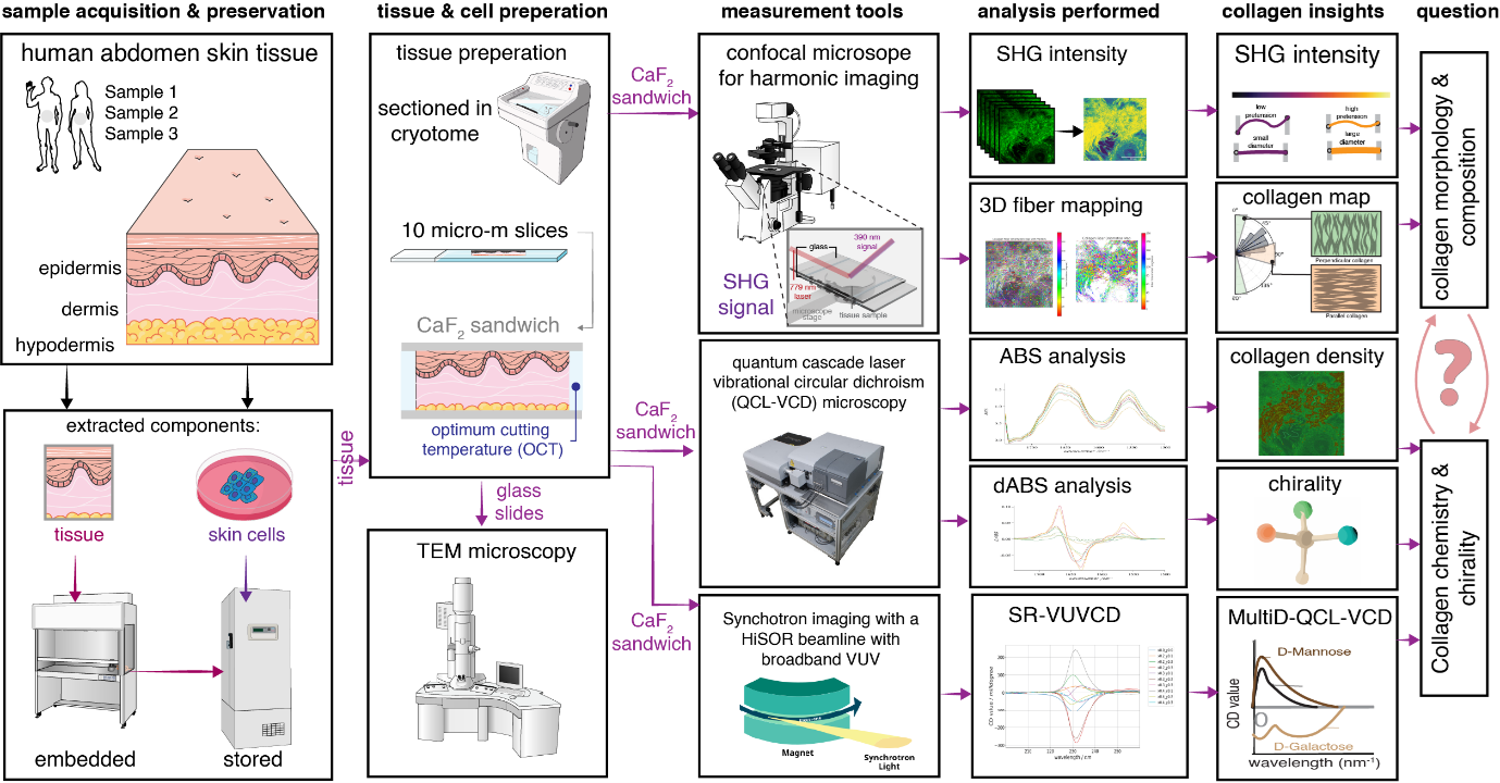
Figure 1: Overview of multifaceted analysis used in this research
Using human skin samples, we evaluated collagen fibrillar structure, density, and supramolecular chirality within the same sample by combining MultiD-QCL-VCD, SHG/confocal laser microscopy, and SR-VUVCD.
Glossary
(*1) Collagen: A protein found in large amounts in skin, bones, tendons, etc. It plays a role in maintaining the firmness and elasticity of the skin.
(*2) WPI-SKCM²: Hiroshima University International Institute for Sustainability with Knotted Chiral Meta Matter. Established after being selected as part of the Ministry of Education, Culture, Sports, Science and Technology's "World Top Level Research Center Program (WPI)" in 2020. An international research center that focuses on substances and structures with knots and chirality, and conducts research across the fields of life, materials, and mathematics.
(*3) Dermis: The tissue beneath the surface of the skin (epidermis) that contains a lot of collagen and supports the strength and elasticity of the skin.
(*4) Chirality: Chirality is the property that the shape reflected in a mirror cannot be superimposed with the original shape, like the left and right hands. For example, if you look at your left hand in a mirror, it will look the same as your right hand.
(*5) Supramolecular chirality: Refers to the chiral order that appears not in a single molecule but in a structure formed by aggregation of multiple molecules. In this study, we focused on the chiral structural order when collagen molecules aggregate to form fibers and networks.
(*6) SHG: Second harmonic generation. A nonlinear optical method for observing regular structures such as collagen fibers without staining.
(*7) SR-VUVCD: Synchrotron radiation vacuum ultraviolet circular dichroism spectroscopy. By measuring circular dichroism in the vacuum ultraviolet region at each position on the sample, collagen triple helix structure and supramolecular chirality were evaluated with high sensitivity without staining.
(*8) MultiD-QCL-VCD: Multidimensional vibrational circular dichroism spectroscopy using a quantum cascade laser. In the infrared region, the molecular structure and supramolecular chirality of collagen were spatially evaluated without staining by mapping and measuring signals originating mainly from the amide I band of collagen (around 1650 cm⁻¹) on the sample.
(*9) Confocal laser microscopy: An imaging method that combines high resolution that exceeds the diffraction limit in the horizontal direction of the sample and spatial resolution in the depth direction. It was used to analyze the distribution, orientation, and network structure of collagen fibers.
Paper information
Publication journal name: ACS Nano (American Chemical Society)
Posting date: July 16, 2026 (Thursday) 13:00 (Japan time)
Paper title: Correlative Multimodal Framework Reveals Supramolecular Chirality Loss Preceding Fibrillar Rarefaction in Dermal Collagen
Author: Ali Haider, Yusuke Kochi, Andrew K. Schulz, Kuya Aoyama, Aiko Sada, Hisako Sato, Elisabetta Matsumoto, Malcolm Kadodwala, Koichi Matsuo, Katsuya Inoue
DOI: 10.1021/acsnano.6c06602
- Press release materials (1.05 MB)
- Article publication journal (ACS Nano)
- Hiroshima University Researcher Guidebook (Professor Katsuya Inoue)
Hiroshima University Graduate School of Advanced Science and Engineering Chemistry Program
Chiral Knot International Research Institute for Supermaterials Contributing to Sustainability
Professor Katsuya Inoue
TEL: 082-424-7416
E-mail: kxi*hiroshima-u.ac.jp
■Matters related to reporting
Hiroshima University Public Relations Office
TEL: 082-424-4518 FAX: 082-424-6040
E-mail: koho*hiroshima-u.ac.jp
(Please replace * with half-width @)